技术平台系列培训2024年第二十二期:病理高阶培训通知
病理高阶培训通知
第一部分:
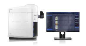
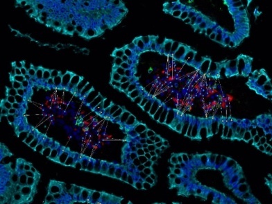
研究所新购置感染性病理切片高分辨全景成像系统,包括蔡司全自动数字玻片扫描系统Axioscan7和艾克发多靶点7色试剂盒,可快速可靠地获取荧光、明场图像。该设备以26 mm × 76mm的标准格式对多达100 张玻片进行自动化扫描,实现单次8色成像。
该系统可应用于:
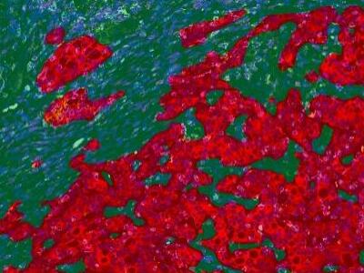
1. 多色荧光解决方案 – 识别不同组织类型的表型,解决肿瘤免疫对多色荧光快速成像的需求。
2. 荧光原位杂交(FISH) – 在基因组中使用多通道荧光和景深延伸来探知在染色体组中单个序列的拷贝数量。
3. 组织芯片技术(TMA) – 可靠的样品检测和稳定的扫描过程,有效的利用试剂以及样品组织;
4. 老年痴呆症与其他相关性疾病,建立对淀粉样沉积物的分析模型。
5. 癌症研究 – 使用虚拟显微技术以出色的荧光虚拟切片来研究癌症发病的根本。
第二部分:
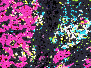
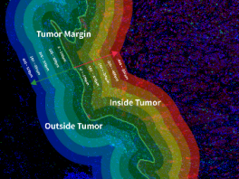
HALO数字病理图像分析平台可用于全组织切片病理图像的精准定量分析,为H&E、IHC、mIF及特殊染色等组织样本提供了多场景分析需求,包括组织分割、组织面积定量、细胞定量以及组织原位的空间位置分析等多种功能。凭借其无与伦比的易用性和灵活性,高度的开放性、全面的分析功能以及高性能的深度学习网络模型,已广泛应用于神经科学,代谢组学,肿瘤学,毒理病理学的高通量,全切片图像的定量研究,满足不同领域,不同学科,从分子到蛋白以及组织层面的多重分析需求。
|
组织分型(肿瘤/间质) |
mIHC免疫细胞亚群定量 |
免疫细胞浸润程度分析 |
细胞间空间邻近距离评估 |
|
|
|
|
|
培训日程:
2024.10.15(星期二)
9:30-12:00,A201,Axioscan7+染色试剂 理论培训(不限制)
13.30-16.00,A201,Halo软件理论和演示培训(不限制)
2024.10.16(星期三)
9:30-17:30,A306,Axioscan7分组上机操作(限名额)
9:30-17:30,多色染色制样培训(限名额)
2024.10.17(星期四)
9:30-15:00,多色染色制样培训(限名额)
课程讲师:蔡司显微镜,产品工程师,郑楠;
艾克发生物,应用科学家,许梦楠;
HALO,应用工程师,赵永田。
请以课题组为单位集中报名至jshao@siii.cas.cn。由于场地限制,将根据报名情况分组上机培训。
报名截止时间:2024.10.11,17:00。
联系人:邵将,54923036。
公共技术中心
公共仪器与测试分析平台
Training Notice for High-resolution Panoramic Imaging System and HALO
PART 1:
The high-resolution panoramic imaging system, including Alphaxbio multi-target 7-color kit and Zeiss Axioscan7 fully automated digital slide scanner, this system can quickly and reliably acquire fluorescence and bright-field images for automated scanning of up to 100 slides in the standard 26 mm × 76 mm format. Optimal spectral separation of up to 8 fluorescent dyes.
It can be used for:
1. Multicolor fluorescent solution, identifying phenotypes of different tissue types, meeting the needs of tumor immunity for rapid imaging with multicolor fluorescence.
2. Fluorescence in-situ hybridization (FISH) - Determining the number of single sequence copies in the genome.
3. Tissue microarrays (TMA) - Resource-friendly use of reagents and tissues with increased throughput.
4. Research on Alzheimer pathogenesis and other age-related diseases - developing analytical models for amyloid deposition (plaques).
5. Cancer research - Researching the basics of cancer.

PART 2:
HALO digital pathology image analysis platform can be used for accurate quantitative analysis of whole slide images, providing multi-scenario analysis needs for tissue samples such as H&E, IHC, mIF and special staining, including tissue segmentation, area quantification, cell quantification, and spatial analysis of tissues in situ and other functions. With its unparalleled ease of use and flexibility, high openness, comprehensive analysis functions, and high-performance deep learning network models, it has been widely used in high-throughput, whole slide images quantitative research in neuroscience, metabolomics, oncology, and toxicological pathology to meet the needs of multiple analyses in different fields, disciplines, and at the level of molecules to proteins and tissues.
|
Tumor/Stromal Tissue Segmentation |
mIHC Immune Cell Subset Quantification |
Analysis of immune cell infiltration level |
Assessment of intercellular space proximity |
|
|
|
|
|
Training time and location:
2024.10.15 (Tuesday)
9:30-12:00, A201, Axioscan7+ Alphaxbio kit Theoretical Training
13.30-16.00, A201, Halo Theoretical and Presentation Training
2024.10.16 (Wednesday)
9:30-17:30, A306, Axioscan7 Operation training (Limited quota)
9:30-17:30, Alphaxbio Operation training (Limited quota)
2024.10.17 (Thursday)
9:30-15:00, Alphaxbio Operation training (Limited quota)
Speaker: ZHENG Nan, ZEISS.
XU Mengnan, Alphaxbio.
ZHAO Yongtian, HALO.
Registration method: Please register as an unit of the research group to jshao@siii.cas.cn . Due to venue limitations, training will be grouped based on the number of applicants.
Registration deadline: 17:00, Oct 11, 2024.
Contact: SHAO Jiang, 54923036.
Institutional Center for Shared Technologies and Facilities
Analytical Core Facility
附件下载: